
In This Issue
Longevity
-
 The CALERIE Receipts: A New Genomic Window Into How Caloric Restriction Shapes Human Aging
Researchers have released a multi-tissue genomic dataset from the only long-term randomized trial of caloric restriction in healthy adults. It won't tell you what to eat tomorrow — but it may finally show how eating less rewires the biology of getting older.
The CALERIE Receipts: A New Genomic Window Into How Caloric Restriction Shapes Human Aging
Researchers have released a multi-tissue genomic dataset from the only long-term randomized trial of caloric restriction in healthy adults. It won't tell you what to eat tomorrow — but it may finally show how eating less rewires the biology of getting older.
-
 Bones, Muscles, and the Frailty Cliff: What a New Aging Study Really Says
A long-running Taiwanese cohort took a closer look at osteosarcopenia — losing bone and muscle at the same time — and the findings are more nuanced than the headlines suggest.
Bones, Muscles, and the Frailty Cliff: What a New Aging Study Really Says
A long-running Taiwanese cohort took a closer look at osteosarcopenia — losing bone and muscle at the same time — and the findings are more nuanced than the headlines suggest.


The CALERIE Receipts: A New Genomic Window Into How Caloric Restriction Shapes Human Aging
Researchers have released a multi-tissue genomic dataset from the only long-term randomized trial of caloric restriction in healthy adults. It won't tell you what to eat tomorrow — but it may finally show how eating less rewires the biology of getting older.
For decades, the idea that eating a little less might help us age a little better has lived in a strange place — somewhere between laboratory fact and dinner-table folklore. Worms live longer on lean rations. So do flies, mice, and at least some monkeys. The harder question, the one that matters to a 65-year-old man trying to stay strong and sharp into his eighties, has always been whether any of that translates to humans. Now a group of researchers has done something quieter than a breakthrough but, in the long run, possibly more useful. They have opened the molecular books.
The work in question is a new genomic data resource drawn from CALERIE — the Comprehensive Assessment of Long-term Effects of Reducing Intake of Energy — the only long-term randomized controlled trial of caloric restriction (CR) ever conducted in healthy, non-obese adults. Writing in Nature Aging, the team behind the resource has released a curated set of genomic data from 218 trial participants, drawn from blood, skeletal muscle and adipose tissue, and sampled at three points across the study. It is not a new finding about caloric restriction so much as a new lens through which the old findings can finally be examined in detail.
That distinction matters. Caloric restriction has been studied in model organisms for nearly a century, and the headline message — that animals fed somewhat less, but not starved, often live longer and healthier lives — has been remarkably durable. The trouble is that humans are not mice. Our lives are longer, messier and harder to randomize. CALERIE, which asked healthy adults to cut their calories by a target of 25 percent for two years, was the field's most serious attempt to bring that question into a rigorous human setting. Earlier reports from the trial broadly supported the pattern seen in animals: signs that the biology of aging itself was being nudged, modestly, in a favorable direction.
What the resource actually contains
The new release is, in plain terms, a paper trail. According to the published description, the resource bundles whole-genome single-nucleotide polymorphism genotypes together with three-timepoint longitudinal DNA methylation, messenger RNA and small RNA datasets. Those measurements were taken from blood, skeletal muscle and adipose tissue samples, for a total of 2,327 samples across the 218 participants. The data are housed at the Aging Research Biobank, available to qualified researchers.
Translated out of the jargon: the team has preserved not just what genes participants carry, but how those genes were being read, regulated and silenced before, during and after a sustained period of eating less. That is the kind of dataset that lets other scientists go back and ask new questions — about specific aging pathways, about why some people responded more than others, about which tissues mattered most — without having to run another two-year human trial to do it.

The CALERIE biospecimens, banked years ago, are now yielding a second life as a multi-omics resource.
Why a dataset is news
It is fair to ask why the release of a database, rather than a fresh result, deserves a reader's attention. The answer is that in geroscience — the science of aging itself — the bottleneck has rarely been ideas. It has been good human data. CALERIE is the rare trial that combined random assignment, a serious intervention, healthy non-obese volunteers, and the patience to follow them for years. The samples it banked are, in a quiet way, irreplaceable. You cannot easily rerun that experiment.
By assembling those samples into a multi-tissue, multi-omics, longitudinal resource and making it available to the broader research community, the team has done something closer to building a library than publishing a paper. The authors describe it explicitly as a resource with great potential to advance translational geroscience — careful language, and the right kind. The promise is not that we now know more about caloric restriction. The promise is that more people, asking better questions, can now find out.
The bottleneck in geroscience has rarely been ideas. It has been good human data.
What this is — and isn't
A few honest caveats are in order, because the gap between a promising dataset and a prescription you would act on is wide. CALERIE enrolled healthy, non-obese adults, most of them well short of the age at which the diseases of aging start to bite hardest. A 25 percent calorie cut, sustained for two years, is a serious undertaking; the participants did not hit that full target on average, and the trial was not designed to test extreme regimens, fasting protocols or the various commercial diets that borrow CR's vocabulary. The earlier human findings from CALERIE, while encouraging, suggest a modest nudge to the biology of aging, not a reversal of it. The new resource expands what can be studied. It does not, by itself, change what is known.
For a reader in his sixties or seventies, the practical takeaway is therefore narrow but real. The science of how eating patterns shape the biology of aging in humans is finally getting the kind of raw material it has long needed. Over the next several years, expect a wave of reanalyses examining how CR affected DNA methylation patterns linked to biological age, gene expression in muscle and fat, and the small-RNA signaling that helps tissues talk to one another. Some of that work will sharpen the case for moderate restraint at the table. Some of it will complicate it. Both outcomes are useful.

- What's new: A curated genomic data resource from the CALERIE trial, covering 218 participants and 2,327 samples, is now available to researchers.
- What's in it: Genotypes plus longitudinal DNA methylation, mRNA and small-RNA data from blood, skeletal muscle and adipose tissue.
- Why it matters: CALERIE is the only long-term randomized trial of caloric restriction in healthy, non-obese adults — its samples are effectively irreplaceable.
- What it isn't: A new finding about caloric restriction, a diet plan, or evidence that aggressive calorie cutting is safe or beneficial for older adults.
- What to watch: Follow-on analyses of CR's effects on epigenetic aging clocks and tissue-specific aging pathways over the next several years.
- For readers: Discuss any substantial change in eating patterns with your clinician, particularly if you are managing weight, muscle mass or chronic conditions.
The longer view here is the one worth holding. For most of the modern era, the study of human aging has been forced to rely on observation: who lived long, who didn't, and what they happened to eat or do along the way. A randomized trial with banked tissues and a fully released genomic resource is a different kind of evidence — slower to arrive, harder to fake, and far more useful to the next generation of researchers. The CALERIE participants, by signing up for two of the most disciplined years of their lives, may end up contributing more to what we know about human aging than anyone fully appreciated at the time. That is a quiet sort of legacy, and a good one.
Frequently asked questions
What is CALERIE, and why is it considered significant?
CALERIE — the Comprehensive Assessment of Long-term Effects of Reducing Intake of Energy — is the only long-term randomized controlled trial of caloric restriction ever conducted in healthy, non-obese adults. It asked participants to cut their calories by a target of 25 percent for two years, making it the field's most serious attempt to bring caloric restriction research into a rigorous human setting.
What exactly is in the new genomic data resource released from CALERIE?
The resource includes whole-genome single-nucleotide polymorphism genotypes along with longitudinal DNA methylation, messenger RNA, and small RNA datasets drawn from blood, skeletal muscle, and adipose tissue. In total, it covers 218 trial participants and 2,327 samples collected at three timepoints across the study, and is housed at the Aging Research Biobank.
Does this new resource prove that eating less extends human lifespan?
No — the authors describe it as a resource rather than a new finding, and the article is explicit that it does not by itself change what is currently known about caloric restriction. Earlier CALERIE results suggested only a modest nudge to the biology of aging, not a reversal of it.
Who were the participants in CALERIE, and did they fully meet the calorie-reduction target?
Participants were healthy, non-obese adults, most of them well short of the age at which the diseases of aging start to bite hardest. On average, they did not hit the full 25 percent calorie-reduction target.
Why should older adults be cautious about applying this research to their own diets?
The article notes that caloric restriction in healthy younger adults is not the same intervention as eating less in your seventies. In older men especially, unintended weight loss and reduced protein intake can erode muscle mass and bone density, which the article describes as the very things that underwrite independence.
Sources
- The CALERIE Genomic Data Resource. — Nature aging

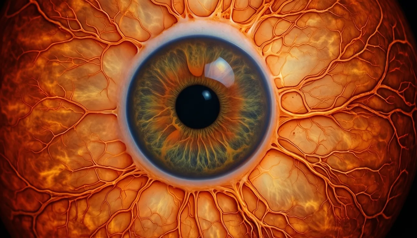
Your Retinas May Be Aging Faster Than You Are — and Multimorbidity Follows
A large deep-learning study suggests the gap between your retina's biological age and your calendar age tracks with chronic disease risk. The eye, it turns out, may be a useful longevity dashboard.
Your calendar age is a stubborn number. Your biology is more negotiable — and increasingly, more measurable. Among the newest candidates for a practical longevity readout is the retina, the thin sheet of neural tissue at the back of the eye that doubles as a window onto the body's small vessels and nerves. A deep-learning analysis of 45,436 middle-aged and older adults, published in GeroScience, reports that the gap between a person's retina-predicted age and their actual age is associated with both current chronic-disease burden and the future onset of multimorbidity. The finding is not a verdict on any one reader's health. It is, however, a useful reframing: the eye may be one of the more honest mirrors we have.
- The metric: 'Retinal age gap' is how much older (or younger) a deep-learning model judges your retina to be versus your calendar age.
- The signal: In a study of 45,436 adults, people with one or two-plus age-related conditions had measurably larger retinal age gaps at baseline than those with none.
- The forecast: Each 5-year increase in retinal age gap was linked to an 8% higher risk of developing multimorbidity over a median 11.4 years of follow-up.
- The caveat: This is an association in one large cohort, not a clinical test you can act on yet. Evidence strength is moderate.
- The use case: Treat retinal imaging as one possible future input to whole-person risk — not a diagnosis, and not a reason to chase a number.
Why the eye keeps showing up in longevity research
The retina is the only place in the body where clinicians can directly observe small arteries, veins and nerve fibers without cutting anything open. Those microvessels respond to the same pressures, glucose excursions and inflammatory traffic that age the rest of the cardiovascular and neurological system. That is why retinal photographs have quietly become a favored training set for biological-age models: the images are standardized, abundant, and tied to long follow-up data in biobanks.
The GeroScience team applied a previously developed deep-learning model that estimates 'retinal age' from a fundus photograph, then subtracted each participant's calendar age to produce a retinal age gap. They then asked a deceptively simple question: does that gap track with how many chronic diseases a person has, and with how many they go on to develop? In the fully adjusted analysis, the answer was yes on both counts.
A standard fundus image. The same photographs that screen for diabetic eye disease are now training grounds for biological-age models.
What the numbers actually say
At baseline, participants who already carried one age-related condition had a retinal age gap roughly 0.20 years larger than peers with none; those with multimorbidity — two or more conditions — had a gap about 0.25 years larger. Both differences were statistically significant in the fully adjusted model, with confidence intervals that excluded zero. These are population-level averages, not personal verdicts. But they are consistent with the idea that disease burden and retinal aging move together.
The prospective piece is the more interesting one. Over a median follow-up of 11.38 years, 3,607 participants — 17.3% of the at-risk group — developed multimorbidity. Each five-year increment in baseline retinal age gap was independently associated with an 8% higher hazard of incident multimorbidity (HR 1.08, 95% CI 1.02–1.14). 'Independently' is doing real work in that sentence: the association held after adjustment for the usual suspects, which is what elevates this from curiosity to signal.
The retina is the only place in the body where a clinician can directly observe small vessels and nerves without an incision. It was always going to end up on the longevity dashboard.
What this is not
This is one large observational cohort with a single retinal-age model and a single composite outcome. It does not show that intervening on the retina — or on anything that nudges the gap — changes disease trajectories. It does not establish that retinal age gap outperforms simpler clinical inputs like blood pressure, HbA1c or a lipid panel. And it cannot tell an individual reader whether their own retina is aging quickly; that would require the model, the image and a clinical context none of us can self-administer.
The evidence rating here is moderate for good reasons. The sample is large and the follow-up long, which is rare and valuable. But the model is one of several in the field, and external validation across populations, cameras and ethnicities remains an open question. Effect sizes are meaningful at population scale and modest at the individual level — an 8% hazard increase per five-year gap is a signal, not a sentence.
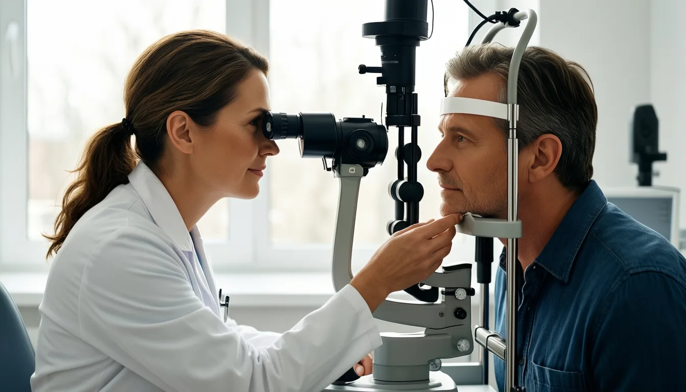
Fundus imaging takes seconds and no dilation in many modern devices. The bottleneck is not the camera — it is the interpretation layer behind it.
How to think about it on a packed calendar
For the executive reader, the practical takeaway is restraint. You do not need a retinal age gap reading to act on what this study reinforces: the same metabolic and vascular forces that age the rest of you also age your retina, and they are largely the levers you already know — sleep, movement, blood pressure, glucose, lipids, stress load. Retinal imaging may eventually sharpen those conversations with a clinician. It does not replace them.
If you already see an optometrist or ophthalmologist annually, ask whether they retain your fundus images and whether their device captures them in a format compatible with future analysis. That is a quiet, no-cost form of optionality. If you are being worked up for cardiometabolic risk, mention that retinal microvascular signals are an active area of research; it may inform how your clinician interprets a borderline finding. Beyond that, the honest advice is to wait for validation and not to chase a number that is not yet yours to chase.
The deeper story the GeroScience analysis tells is not about eyes. It is about the slow normalization of biological age as a clinical concept — a shift from treating chronological age as destiny to treating it as one variable among many. The retina is a convenient proxy because it is photographable, standardized and richly vascular. Other organs will get their own clocks. The reader's job, for now, is to recognize the direction of travel without overreacting to any single instrument on the dashboard.
Frequently asked questions
What exactly is a 'retinal age gap'?
A retinal age gap is the difference between the age a deep-learning model estimates your retina to be — based on a fundus photograph — and your actual calendar age. A positive gap means your retina appears older than you are; a negative gap means it appears younger.
What did the study find about retinal age gap and future disease risk?
Over a median follow-up of 11.38 years, each five-year increase in baseline retinal age gap was associated with an 8% higher hazard of developing multimorbidity. In that period, 17.3% of the at-risk group went on to develop two or more age-related chronic conditions.
Why do researchers use the retina to study biological aging?
The retina is the only place in the body where small arteries, veins, and nerve fibers can be directly observed without any incision. Those microvessels respond to the same pressures, glucose changes, and inflammation that age the cardiovascular and neurological systems, making retinal photographs a useful training set for biological-age models.
Is this a test I can take now to find out how fast my retina is aging?
No — the article describes this as an association from one large observational cohort, not a clinical test you can act on yet. External validation across different cameras, populations, and ethnicities is still an open question, and the evidence strength is rated as moderate.
What practical step, if any, does the article suggest regarding retinal imaging?
The article suggests asking your optometrist or ophthalmologist whether they retain your fundus images and whether their device captures them in a format compatible with future analysis — described as a quiet, no-cost form of optionality. Beyond that, it advises waiting for further validation rather than trying to chase a retinal age gap number.
Sources

Bones, Muscles, and the Frailty Cliff: What a New Aging Study Really Says
A long-running Taiwanese cohort took a closer look at osteosarcopenia — losing bone and muscle at the same time — and the findings are more nuanced than the headlines suggest.
Here's the obvious beginner question I had to ask my editor before writing this: if bones get thinner with age and muscles get smaller with age, what happens when both go at once? There's a name for it — osteosarcopenia — and longevity researchers think it might be one of the earliest warning signs that someone is drifting toward frailty. A new follow-up of a big Taiwanese aging study just took a careful look. The story it tells is real, but quieter than the buzzy headlines.
Let's gloss the jargon first. Sarcopenia is the age-related loss of muscle mass and strength. Osteopenia and osteoporosis are the stages of thinning bone. Osteosarcopenia is just the unhappy overlap — you've got both. And frailty, in research-speak, isn't vague "feeling old." It's a specific clinical syndrome (the Fried criteria), built from things like unintentional weight loss, exhaustion, weak grip, slow walking, and low activity.
The hypothesis researchers have been chasing is intuitive: if your scaffolding (bone) and your engine (muscle) are both weakening, frailty is the destination. A 2025 analysis published in Archives of Gerontology and Geriatrics set out to test exactly that, using one of Asia's most-watched aging cohorts.
What the I-Lan study actually did
The I-Lan Longitudinal Aging Study follow-up started with 1,779 community-dwelling adults aged 50 and up in Taiwan, and checked back in with 998 of them eight years later. At baseline, the researchers measured bone status (using WHO definitions) and sarcopenia (using the Asian Working Group definition). At follow-up, they scored frailty using the Fried criteria. Then they asked: did people with osteosarcopenia at the start end up frail eight years later, more than everyone else?
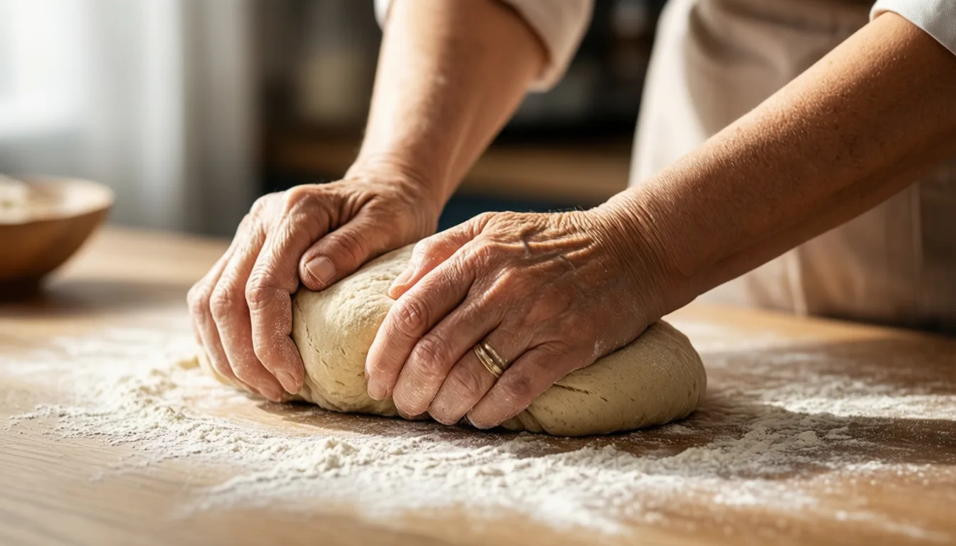
Grip strength is one of the Fried frailty criteria — which is why everyday loads (groceries, dough, garden tools) double as low-key training.
The finding everyone shares — and the finding they skip
Here's the part that's been getting passed around: at baseline, osteosarcopenia was dramatically more common in people who were already frail (27.5%) than in those who were pre-frail (10.8%) or non-frail (0%), according to the I-Lan analysis. That's a striking snapshot. Bone-plus-muscle loss clearly travels with frailty.
But — and this is the part that matters for a longevity reader — when the authors asked whether osteosarcopenia at baseline predicted who became frail eight years later, the association did not reach statistical significance. The odds ratio pointed in the expected direction (OR 2.67), but the confidence interval crossed 1 (95% CI 0.85–8.40, p = 0.094). The same was true for sarcopenia, osteopenia, and osteoporosis on their own. The authors' own conclusion is appropriately measured: neither osteosarcopenia nor its components was significantly associated with frailty risk at eight years in this cohort.
So what do we actually have? A robust cross-sectional link, and a longitudinal signal that is suggestive but not proven. That's why this piece is labeled moderate evidence and not strong. It's a real lead — not a verdict.
Osteosarcopenia clearly travels with frailty. Whether it drives frailty years in advance is still an open question.
Why this still matters in your 40s and 50s
Even with a cautious read of the data, the practical takeaway doesn't really change. The I-Lan cohort had a mean baseline age of 63.9 — meaning the bone and muscle people brought into their 60s was largely built (or not built) in the decades before. Frailty is not a switch that flips at 75. It's a slow drift, and the inputs that bend the curve — resistance training, enough protein, staying active outside of "workouts" — are the same inputs that look protective across nearly every aging study we have.
The smart-friend version: you're not training today because you'll be frail tomorrow. You're training because the version of you that's 70 will be working with whatever bone density and muscle mass you bank now. Lifting something heavy-ish a few times a week, getting protein at every meal, and walking a lot is the unsexy core. Everything else is seasoning.
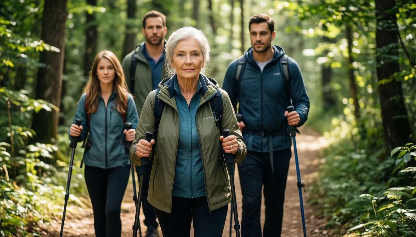
Community-dwelling older adults — the population the I-Lan study followed — are exactly who longevity guidance is built around.
- Osteosarcopenia is a real concept. It's the overlap of weak bones and shrinking muscle — and at any given moment, it tracks closely with frailty.
- The new I-Lan follow-up is more cautious than the headlines. At 8 years, osteosarcopenia did not significantly predict who became frail, though the trend pointed that way.
- Cross-sectional ≠ causal. Seeing two things together in older adults today doesn't prove one caused the other tomorrow.
- The action items don't change. Resistance training and adequate protein remain the best-supported levers for preserving both bone and muscle.
- Frailty is built decades early. Most participants were already in their 60s at baseline — the time to invest is well before then.
- This is education, not prescription. Talk to a clinician before starting a new training program or changing your diet, especially if you've had fractures or balance issues.
What I'll be watching next
The I-Lan authors are clear that data on osteosarcopenia and frailty risk is still scarce, and their own follow-up couldn't seal the case. The most useful next chapter would be larger, longer cohorts that catch people before any bone or muscle loss is measurable — and that test whether training and nutrition interventions actually bend the frailty curve, not just the lab values. Until then, the honest framing is the one a good clinician would give you: the link is plausible, the snapshot is suggestive, and the basics are worth doing anyway.
Frequently asked questions
What is osteosarcopenia?
Osteosarcopenia is the overlap of two age-related conditions: sarcopenia, which is the loss of muscle mass and strength, and osteopenia or osteoporosis, which are the stages of thinning bone. Longevity researchers think it may be one of the earliest warning signs that someone is drifting toward frailty.
How did the I-Lan study define frailty?
The study used the Fried criteria, a specific clinical framework built from measurable factors including unintentional weight loss, exhaustion, weak grip strength, slow walking, and low physical activity. In research terms, frailty is not a vague sense of feeling old but a defined clinical syndrome.
Did the study prove that osteosarcopenia causes frailty years later?
No. When researchers asked whether osteosarcopenia at baseline predicted who became frail eight years later, the association did not reach statistical significance — the confidence interval crossed 1 and the p-value was 0.094. The authors' own conclusion is that neither osteosarcopenia nor its components was significantly associated with frailty risk at eight years in this cohort.
How common was osteosarcopenia among people who were already frail at the start of the study?
At baseline, osteosarcopenia was present in 27.5% of participants who were already frail, compared to 10.8% of those who were pre-frail and 0% of those who were non-frail. The article describes this as a striking cross-sectional snapshot showing that bone-plus-muscle loss clearly travels with frailty.
Why does the article say this research still matters for people in their 40s and 50s?
The mean baseline age of the I-Lan cohort was 63.9, meaning the bone and muscle participants brought into their 60s was largely built — or not built — in the decades before. The article frames frailty not as a switch that flips at 75 but as a slow drift, and notes that the inputs most likely to help — resistance training, adequate protein, and staying active — appear protective across nearly every aging study available.
Sources
- Osteosarcopenia and frailty risk in community-dwelling older adults: A follow-up of the I-Lan Longitudinal Aging Study. — Archives of gerontology and geriatrics