
In This Issue
Longevity
-
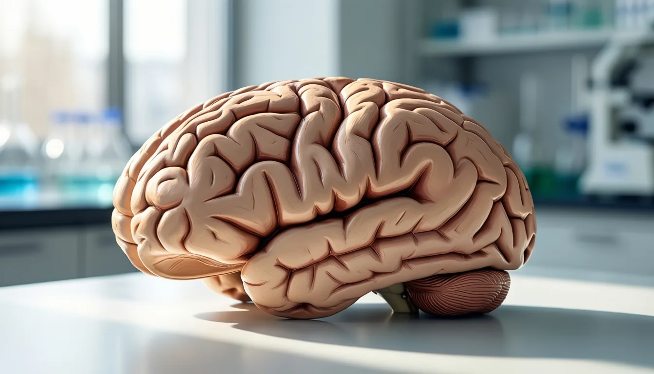
 Reading Organ Age in a Drop of Blood: What 44,000 Plasma Proteomes Tell Us About Living Longer
A massive new study suggests your body doesn't age all at once — and a single blood draw might one day map which organs are racing ahead.
Reading Organ Age in a Drop of Blood: What 44,000 Plasma Proteomes Tell Us About Living Longer
A massive new study suggests your body doesn't age all at once — and a single blood draw might one day map which organs are racing ahead.
-
 Why Your Biological Age Score Might Be Wrong
A new GeroScience analysis argues the machine-learning clocks behind today's biological-age tests optimize for math, not biology — and miss inflammation in the process.
Why Your Biological Age Score Might Be Wrong
A new GeroScience analysis argues the machine-learning clocks behind today's biological-age tests optimize for math, not biology — and miss inflammation in the process.
-
 Measuring Age Itself: The Quiet Revolution in How We Quantify Getting Older
Two new papers — a framework for what a real aging biomarker must do, and a label-free way to read it inside mitochondria — suggest biological age is finally becoming measurable science.
Measuring Age Itself: The Quiet Revolution in How We Quantify Getting Older
Two new papers — a framework for what a real aging biomarker must do, and a label-free way to read it inside mitochondria — suggest biological age is finally becoming measurable science.
-
 What Companion Dogs Are Teaching Us About Human Aging
The Dog Aging Project's Precision Cohort turns 1,000 family pets into a living laboratory for geroscience — and a useful mirror for the rest of us.
What Companion Dogs Are Teaching Us About Human Aging
The Dog Aging Project's Precision Cohort turns 1,000 family pets into a living laboratory for geroscience — and a useful mirror for the rest of us.


Reading Organ Age in a Drop of Blood: What 44,000 Plasma Proteomes Tell Us About Living Longer
A massive new study suggests your body doesn't age all at once — and a single blood draw might one day map which organs are racing ahead.
Here's a thought that rearranged my week: the number on your birthday cake might be the least interesting age you have. Scientists are starting to talk about your organ ages — plural — because your heart, your brain, your immune system, and eight other organs may each be running on their own clocks. And a recent analysis suggests we can read those clocks in something as ordinary as a vial of blood.
The study, published in Nature Medicine, looked at plasma proteins — the tiny molecular messengers floating around in the liquid part of your blood — from 44,498 people in the UK Biobank. Researchers measured almost 3,000 different proteins per person and used them to estimate the biological age of 11 separate organs. Quick gloss: "biological age" just means how old a tissue looks on the inside, based on its molecular wear and tear, regardless of the candles on the cake.
The headline finding is that those organ ages aren't just a party trick. They forecast real disease — heart failure, chronic obstructive pulmonary disease, type 2 diabetes, Alzheimer's — across a follow-up window stretching up to 17 years. That's the part that made me sit up. We're not talking about predicting next year's flu. We're talking about a blood draw today hinting at what might show up two decades from now.
The brain clock, and why it's the loudest one
Of all 11 organs, two kept stealing the spotlight: the brain and the immune system. People whose plasma proteins suggested an unusually aged brain had a risk of Alzheimer's disease roughly three times higher than average — a risk on par with carrying one copy of APOE4, the best-known genetic risk factor for late-onset Alzheimer's.
Flip it around and the story gets more hopeful. A youthful-looking brain was linked to a risk of Alzheimer's that was roughly a quarter of average — protection comparable to carrying two copies of the protective APOE2 variant, and apparently independent of which APOE genes you actually inherited. In plain language: your genes are not the whole script.
Your body doesn't age on a single clock. It ages on eleven of them — and the brain's tick is the one that echoes loudest.

Eleven organs, eleven clocks: the study estimated biological age organ by organ rather than as a single whole-body number.
Stacking aged organs is where the risk really compounds
Here's the obvious beginner question I had to ask: does it matter if just one organ is running fast, or is it the pile-up that hurts? The data suggests it's very much the pile-up. People with two to four organs flagged as aged had roughly 2.3 times the mortality risk of peers. With five to seven, that climbed to about 4.5 times. Eight or more aged organs pushed it to roughly 8.3 times.
The mirror image is just as striking. A youthful brain alone was linked to a 40% lower mortality risk; a youthful immune system, similar. Having both youthful was associated with a risk cut by more than half. No other organ pairing carried quite the same signal, which is part of why the authors flag the brain and immune system as the most promising places to focus longevity research.

The whole pipeline starts with one small sample. The hard part is teaching the proteins to talk.
What this is — and what it isn't
Time for the careful part. This is one very large observational study. It shows associations, not proof that nudging your proteins around will buy you extra birthdays. The researchers also noted that organ age estimates shifted with lifestyle factors and medications, which is intriguing but doesn't yet tell us which specific changes move which specific clocks, or by how much.
There are also caveats baked into the cohort itself: UK Biobank participants skew healthier, whiter, and more middle-aged than the general population, so we should be cautious about assuming every finding generalizes. And while the modeling is sophisticated, organ-age tests aren't currently a clinical tool you can order at a checkup — they're a research instrument that may, eventually, become one.
- Organs age at different speeds. A single blood draw may, in research settings, estimate the biological age of 11 organs separately.
- Brain and immune age stood out. Both were strongly linked to long-term disease risk and mortality across nearly 45,000 people.
- An aged brain ≈ APOE4-level Alzheimer's risk. A youthful brain showed APOE2-level protection, independent of genotype.
- Risk stacks. Mortality risk rose with the number of organs flagged as aged — modestly at first, sharply past five.
- This is moderate evidence. One large observational study; associations, not proven cause and effect.
- Not a clinical test yet. Talk to a clinician before acting on any longevity or "biological age" product.
The part I keep coming back to is the reframe. For years, the longevity conversation has revolved around a single biological-age number — one score to rule them all. This work suggests that's the wrong resolution. A 50-year-old with a youthful brain and a tired liver is a very different person, healthwise, from a 50-year-old with the reverse. Treating them the same was always a little strange; now we have data hinting at how strange.
None of this is a permission slip to skip sleep, stress, or your next checkup. But it's a genuinely fresh way of thinking about getting older — not as one clock winding down, but as a small orchestra, each instrument keeping its own time. The next question, and it's the one researchers are racing to answer, is which instruments we can actually tune.
Frequently asked questions
How did researchers estimate the biological age of individual organs?
Researchers measured almost 3,000 plasma proteins per person from 44,498 UK Biobank participants and used those proteins to estimate the biological age of 11 separate organs. Biological age refers to how old a tissue looks on the inside based on its molecular wear and tear, regardless of a person's chronological age.
What does an 'aged brain' mean for Alzheimer's risk, according to the study?
People whose plasma proteins suggested an unusually aged brain had an Alzheimer's risk roughly three times higher than average, a level the researchers compared to carrying one copy of APOE4, the best-known genetic risk factor for late-onset Alzheimer's. Conversely, a youthful-looking brain was linked to a risk roughly a quarter of average, comparable to the protection associated with carrying two copies of the APOE2 variant, and apparently independent of which APOE genes a person actually inherited.
Does having just one aged organ raise health risk as much as having several?
The data suggests the pile-up matters most. People with two to four organs flagged as aged had roughly 2.3 times the mortality risk of peers, rising to about 4.5 times with five to seven aged organs, and roughly 8.3 times with eight or more.
Can I order this kind of organ-age test through my doctor today?
No. The organ-specific proteomic profiling described in the study is not currently a clinical tool available at a routine checkup — it is a research instrument that may eventually become one. The article advises treating any direct-to-consumer product that claims to read your organ ages with healthy skepticism and consulting a clinician before making decisions based on such a result.
What are the main limitations of this research?
This is one large observational study, meaning it shows associations rather than proof that changing protein levels will extend life. The UK Biobank cohort also skews healthier, whiter, and more middle-aged than the general population, so the findings may not generalize to everyone.
Sources

Why Your Biological Age Score Might Be Wrong
A new GeroScience analysis argues the machine-learning clocks behind today's biological-age tests optimize for math, not biology — and miss inflammation in the process.
The vial arrives in a tidy box, the instructions promise a number, and the number promises a verdict: you are younger than your birthday, or older, or — most unsettling — somewhere your body has been hiding from you. Consumer biological-age tests have become a fixture of the longevity conversation, sold as a mirror more honest than the calendar. But a 2025 analysis in GeroScience argues that the mirror is warped in a specific, technical way most buyers never hear about: the algorithms behind these scores were built to look smooth on a graph, not to faithfully describe what aging is doing inside a cell.
The paper, by Mei and colleagues, is not a tabloid takedown. It is a careful methodological critique of the machine-learning logic that underpins the most widely used DNA methylation "age clocks" — the kind of model that powers many of the at-home tests now marketed to health-curious consumers. Their central claim is uncomfortable: a clock can be excellent at predicting chronological age and still be a poor instrument for understanding biological aging. The reason is that the math rewards features that move in a clean line from birth toward death, even when the underlying biology does nothing of the sort.
Aging, the authors remind us, is not linear. It has different rhythms in childhood, midlife, and the years after menopause, with abrupt inflection points rather than a tidy slope. When an algorithm is trained to compress that messy reality into a single rising line, it tends to keep the features that cooperate and quietly discard the ones that don't — even if the discarded signals are the biologically interesting ones. The output looks impressive. The interpretability suffers.
The incoherence problem
One of the paper's sharpest observations concerns what the authors call incoherence. In the DNA methylation patterns these clocks read, some sites gain methylation as we age and as disease accumulates; others lose it in the same direction. A biologically faithful model would treat those two groups as opposites. Yet the authors show that major conventional clocks assign positive weights to both kinds of sites, effectively adding signals that should be subtracting from one another. The model still produces a number. It just isn't the number you think it is.
The consequences are not abstract. The same analysis finds that popular clocks are skewed toward leukocyte fractions — heavily influenced by the mix of white blood cells in a sample rather than by aging itself. A score can shift because your immune cell composition shifted that morning, not because anything meaningful changed about your biological trajectory. For a reader weighing whether last quarter's number really reflects last quarter's habits, that is a sobering caveat.

DNA methylation clocks read chemical tags on the genome — but how those readings are combined matters as much as the readings themselves.
What the clocks miss
Perhaps the most striking finding, especially for women navigating the post-menopausal decade, concerns inflammaging — the chronic, low-grade inflammation now considered a hallmark of biological aging and a driver of cardiovascular, metabolic, and cognitive decline. The authors argue that conventional age clocks struggle to detect inflammaging, precisely because the mathematical scaffolding that makes them look accurate also flattens the biological signal that inflammation leaves behind.
When the authors rebuild the models to remove the incoherence — forcing the math to respect the direction biology is actually moving — the picture changes. The corrected models are less skewed toward neutrophils and better at detecting inflammaging. That is not a marketing flourish; it is a methodological proof of concept that the standard approach is leaving important information on the table.
A clock can be excellent at predicting chronological age and still be a poor instrument for understanding biological aging.
How to read your own number
None of this means biological-age testing is meaningless, and the GeroScience authors do not argue that it is. They are clear-eyed about the appeal of a single, legible score, and they sketch a path forward through non-linear machine-learning approaches that may better honor the shape of aging. The point is more measured: today's commercial tests rest on a methodological tradition whose limitations are now being articulated in the peer-reviewed literature, and the informed consumer should know that.
For readers who have already taken a test, the practical reframing is this. A biological-age score is best understood as one signal among many — interesting, not authoritative, and likely sensitive to factors (like the immune cell mix in a given blood draw) that have little to do with how you have been living. A score that drops after a quarter of better sleep and walking is encouraging; a score that jumps after a stressful month is not a diagnosis. Anyone weighing what a result means for their actual health decisions should talk it through with a clinician who can place it alongside the rest of the picture — blood pressure, lipids, inflammatory markers, function, history.
The longevity field is moving quickly, and the candor of this paper is itself a good sign. The same researchers building the next generation of clocks are publicly auditing the last one. That is how a young science matures — not by retreating from the promise of measuring aging, but by being honest about which measurements have earned our trust, and which are still a work in progress.
- The critique is methodological, not commercial. A 2025 GeroScience analysis argues popular DNA methylation clocks optimize for mathematical linearity at the cost of biological meaning.
- Incoherence is the core issue. Major clocks assign positive weights to methylation sites that move in opposite biological directions, muddying what the score represents.
- The numbers can drift for the wrong reasons. Conventional clocks are skewed toward leukocyte fractions, meaning immune cell composition can shift your result.
- Inflammaging is being missed. Standard models struggle to detect chronic low-grade inflammation, one of the most consequential features of aging.
- Treat a score as one signal, not a verdict. Discuss results with a clinician who can interpret them alongside conventional health markers.
- The field is correcting itself. Non-linear and coherence-corrected models are being explored as a more biologically faithful next step.

The behaviors that move biological-age scores — sleep, movement, stress, nutrition — are also the ones with the strongest independent evidence for healthy aging.
Frequently asked questions
Why can my biological age score shift even if my lifestyle hasn't changed?
According to the article, conventional clocks are skewed toward leukocyte fractions, meaning they are heavily influenced by the mix of white blood cells present in a blood sample on a given day. A score can shift because your immune cell composition changed at the time of the draw, not because anything meaningful changed about your biological trajectory.
What is the 'incoherence' problem the researchers identified?
Incoherence refers to the finding that major conventional clocks assign positive weights to methylation sites that move in opposite biological directions — some sites gain methylation with age and disease while others lose it, yet both are treated as additive signals. A biologically faithful model would treat those two groups as opposites, but instead the math combines them, muddying what the final score actually represents.
Why do these clocks struggle to detect inflammaging, and why does that matter?
The article explains that the same mathematical scaffolding that makes conventional clocks appear accurate also flattens the biological signal that inflammation leaves behind. This matters because inflammaging — chronic, low-grade inflammation — is described as a hallmark of biological aging and a driver of cardiovascular, metabolic, and cognitive decline.
Does this analysis mean biological age testing is useless?
The GeroScience authors do not argue that biological age testing is meaningless, and the article takes the same measured position. It describes a score as one signal among many — interesting but not authoritative — and notes that the field is actively exploring non-linear and coherence-corrected models as more biologically faithful alternatives.
How should I interpret a score that went up or down after a big change in my routine?
The article frames a score that drops after a quarter of better sleep and walking as encouraging, while noting that a score that jumps after a stressful month is not a diagnosis. It recommends discussing what a result means with a clinician who can place it alongside other health markers such as blood pressure, lipids, inflammatory markers, function, and personal history.
Sources
- Misalignment of age clocks. — GeroScience

AI at the Dinner Plate: What 'Precision Nutrition' Actually Delivers Today
Algorithms promise to engineer the perfect meal for your face, your physique, and your future. A new scoping review separates the signal from the marketing.
The pitch is irresistible to anyone who treats their body like a project. Upload your bloodwork, strap on a sensor, swab your gut, and let a model spit back the exact bowl of food that will sharpen your jawline, smooth your skin, and stretch your healthspan by a decade. Precision nutrition powered by artificial intelligence has become the glossiest promise in the wellness aisle — and, finally, it has a serious literature behind it worth interrogating. A new scoping review in Advances in Nutrition pulled almost two hundred studies into one room and asked the only question that matters for readers optimizing in real life: what part of this is actually working yet, and what is still a beautifully designed wait.
- The field is exploding, fast. Roughly three-quarters of AI precision-nutrition research has been published since 2020, concentrated on diet-related disease like diabetes and cardiovascular conditions.
- The real wins are narrow. Continuous glucose monitor–guided recommendations and microbiome modeling are the most developed use cases — promising, but still maturing.
- Evidence is uneven. Methods, datasets, and evaluation metrics vary widely across studies, making head-to-head comparisons difficult.
- Diversity is the elephant in the lab. Minority and cultural representation is thin, which limits how well today's models generalize to real eaters.
- Treat outputs as hypotheses, not prescriptions. The smartest move right now is using these tools to ask better questions of a clinician — not to replace one.
What the review actually found
The authors followed a PRISMA-ScR scoping protocol and pulled 198 articles spanning precision nutrition, AI, and natural language processing, then mapped the landscape: where the work is being published, which conditions it targets, what methods it leans on, and — crucially for a category sold on personalization — whether it accounts for the humans it claims to personalize for.
The headline trend is momentum. About 75% of the studies in the review were published from 2020 onward, a surge that tracks with the broader explosion in machine learning tooling and the consumer rise of wearables. The dominant clinical targets are predictable and reasonable: diabetes and cardiovascular disease, with a secondary emphasis on prevention and general health optimization. That is meaningful for the looksmaxing reader, because the same metabolic levers that drive cardiometabolic risk — glycemic control, inflammation, body composition — are the levers that quietly govern skin quality, hair density, sleep architecture, and the soft-tissue contours people obsess over.

CGM-driven recommendations are among the most developed real-world applications mapped in the review.
So where is the work strongest? The review describes a maturing toolkit built around two pillars that any reader who has shopped a wellness app will recognize. The first is AI applied to continuous glucose monitoring data to predict individual glycemic responses to specific meals — the basic premise behind the personalized-blood-sugar category. The second is microbiome modeling, where machine learning tries to translate a stool sample's bacterial signature into food recommendations. Both are real research programs, not vibes. Both are also, in the review's framing, still part of an evolving landscape rather than a settled science.
The dream is a model that knows your dinner before you do. The reality is a research field still learning what 'you' even means in its training data.
The diversity problem nobody markets around
If there is one finding that should reset the way readers shop this category, it is this: the review explicitly flags the need to better integrate minority and cultural perspectives to make AI precision nutrition equitable and effective. Translation for the consumer: a recommendation engine is only as personalized as the people it was trained on. If a model was built largely on one demographic's diets, microbiomes, and glycemic patterns, its confident-sounding output for someone outside that group is closer to an educated guess than a precision call.
This matters double for cultural eating patterns. A model that has barely seen injera, congee, dal, or pozole will struggle to give meaningful guidance to anyone whose plate actually looks like their family's. The review treats this as a research gap to close, not a dealbreaker — but it is the gap most worth keeping in mind when an app tells you, with apparent certainty, which carb to cut.
What this means for the optimizer
For the reader whose health interest skews aesthetic — clearer skin, leaner composition, deeper sleep, better recovery — the practical read is more nuanced than "it works" or "it doesn't." The review's signal is that AI precision nutrition is a legitimately active field with concentrated traction in glycemic and microbiome use cases, particularly aimed at metabolic disease. Those are the same upstream systems that quietly determine how your face looks at 7 a.m. on a Tuesday.
But the methodological variation the review documents — different datasets, different evaluation metrics, different definitions of success — is the kind of heterogeneity that makes it hard to say any one consumer product is doing what its marketing implies. A glucose-prediction app that performs beautifully in one cohort may not generalize cleanly to you. A microbiome recommendation engine may be drawing from a reference population that does not include your ancestry, your diet, or your medication list.
None of this is a reason to dismiss the category. It is a reason to use it the way thoughtful early adopters use any moderately evidenced tool: as a structured way to notice patterns, generate hypotheses, and have a sharper conversation with a clinician who can interpret them against your bloodwork and history. The plate in front of you is still the variable that matters most. The algorithm, for now, is a second opinion worth weighing — not the chef.

Personalization is only as deep as the data behind it — cultural diet patterns remain underrepresented in the literature.
The most honest framing of where this category sits in 2026 is the one the scoping review itself implies: a fast-moving research front with credible early applications, real methodological work still ahead, and a personalization promise that will only get truer as the training data starts to look like the people buying the subscription. For now, the smartest move at the dinner plate is the same one it has always been — eat like someone who plans to be around, and audit the algorithm before you let it audit you.
Frequently asked questions
Which AI precision nutrition applications are the most developed right now?
According to the review, the two strongest use cases are AI applied to continuous glucose monitor data to predict individual blood sugar responses to specific meals, and microbiome modeling that translates a stool sample's bacterial signature into food recommendations. Both are described as real research programs, though still part of an evolving landscape rather than settled science.
Why might an AI nutrition app's recommendations not apply to me?
The review explicitly flags that minority and cultural perspectives are underrepresented in the research, meaning a model built largely on one demographic's diets, microbiomes, and glycemic patterns may produce results that are closer to an educated guess than a precision call for anyone outside that group. Cultural eating patterns — foods like injera, congee, dal, or pozole — remain largely absent from training data.
How should I actually use AI nutrition tools given that the science is still maturing?
The review's framing suggests using these tools as a structured way to notice patterns and generate hypotheses, then bringing that data to a clinician who can interpret it against your bloodwork and history. The article describes the algorithm as a second opinion worth weighing, not a replacement for a clinician.
Why is it hard to know whether a specific precision nutrition product does what it claims?
The review documents wide variation across studies in datasets, evaluation metrics, and definitions of success, which makes head-to-head comparisons difficult. A glucose-prediction app that performs well in one cohort may not generalize to someone with a different ancestry, diet, or medication list.
What health conditions does most AI precision nutrition research focus on?
The dominant clinical targets in the review are diabetes and cardiovascular disease, with a secondary emphasis on prevention and general health optimization. The article notes these same metabolic levers — glycemic control, inflammation, and body composition — also influence factors like skin quality, sleep, and body composition more broadly.
Sources
- A Scoping Review of Artificial Intelligence for Precision Nutrition. — Advances in nutrition (Bethesda, Md.)

Eat Less, Eat Later: What New Research on Caloric Restriction and Time-Restricted Eating Means for the Aging Heart
Two 2025 papers connect how we eat — and when — to the metabolic stress pathways behind a stubborn form of heart failure. The science is promising, the certainty is not.
Somewhere between the third perimenopause article that told me to just eat less and the fourth one that told me to just eat in a window, I started wondering whether anyone could explain what either of those things is actually doing inside the body — and whether the heart, the organ we tend to ignore until it gets loud, cares about the difference. As it turns out, two 2025 papers tried to answer exactly that. Neither is the miracle headline you'll see on Instagram. Both are more interesting than that.
Here's the setup. A form of heart failure called HFpEF — heart failure with preserved ejection fraction — has been quietly becoming one of the defining cardiovascular problems of midlife and beyond, especially for women. The pump still squeezes fine on an echocardiogram. The problem is that the heart muscle has gotten stiff, the metabolism around it has gotten messy, and the usual heart-failure drugs don't move the needle the way they do in other kinds of heart failure. Obesity, insulin resistance, and the cellular wear-and-tear of aging all stack the deck.
That's the backdrop for a new review in Cardiovascular Diabetology, which lays out the case that HFpEF is, at its core, a disease of accelerated metabolic aging — and that caloric restriction (and drugs that mimic its effects) may be one of the few interventions that targets the underlying biology rather than just the symptoms. The authors are careful: this is a review of mechanisms and emerging therapeutics, not a verdict. But the throughline is striking. The same cellular hallmarks that show up in aging tissue generally — inflammation, mitochondrial drift, faulty nutrient sensing — show up amplified in HFpEF hearts.
- HFpEF is metabolic, not just mechanical. A 2025 review frames it as a disease where aging biology and obesity converge on the heart.
- Caloric restriction has biological plausibility — and human signal. Weight-loss strategies, particularly calorie reduction, are described as showing promise in HFpEF patients, though this is not a settled treatment.
- "CR mimetics" are a real research category. Some clinically approved drugs may replicate parts of the calorie-restriction signal without the diet.
- Time-restricted feeding shifts gene expression — tissue by tissue. In aged rats, eating-window changes altered metabolic-pathway genes differently in brain, liver, and muscle.
- Evidence rating: moderate. Mechanistic, preclinical, and review-level data are strong; large human outcome trials in HFpEF are not yet here.
What "caloric restriction" actually does (besides making you hungry)
When researchers talk about caloric restriction, they don't mean starvation and they don't mean a Tuesday salad. They mean a sustained, modest reduction in calories that — in model organisms, reliably, and in humans, more tentatively — bends the curve on biological aging. It nudges nutrient-sensing pathways like mTOR, AMPK, sirtuins, and the PI3K/AKT axis toward a state that looks less like "grow and store" and more like "repair and maintain."
The HFpEF review argues that this matters for the aging heart specifically because the failure mode in HFpEF isn't a blown-out pump — it's a heart that has become metabolically inflexible, surrounded by tissue that's inflamed and insulin-resistant. Calorie reduction, the authors write, appears to mitigate disease progression by acting on those upstream aging mechanisms rather than the downstream cardiac symptoms.
The honest caveat: the gold-standard evidence for caloric restriction extending healthspan still comes from animal studies and small human trials. The HFpEF-specific human data is suggestive, not definitive. This is a moderate-evidence story, not a settled one.
The failure mode in HFpEF isn't a blown-out pump — it's a heart that's become metabolically inflexible.

Researchers are increasingly framing the aging heart as a metabolic organ, not just a mechanical one.
The "mimetic" question — can a pill do what a diet does?
Here's where it gets interesting for anyone who has tried, and not loved, sustained calorie reduction. The review explicitly takes up the question of caloric restriction mimetics — compounds, including some already in clinical use, that may reproduce parts of the calorie-restriction signal pharmacologically. The thesis is that these could sidestep the very real adherence problem of asking aging humans to permanently undereat.
I want to be careful here, because this is precisely the territory where supplement marketers love to plant a flag. The review treats mimetics as a serious research direction with a credible mechanistic rationale. It does not declare them ready for prime time as HFpEF therapy. Several candidates are repurposed metabolic drugs you may already know about. None of them is the resveratrol gummy on your friend's counter.
What about when you eat?
The second 2025 paper — from GeroScience — moves the conversation from how much to when. Researchers fed aged rats one of three diets from mid-life onward: ad libitum (anything, anytime), time-restricted feeding with normal macros (cTRF), or time-restricted feeding with ketogenic macros (kTRF). Then they looked at gene expression along the PI3K/AKT metabolic pathway across brain, liver, and skeletal muscle.
Two things stand out. First, the effects were tissue-specific. In the brain — specifically the CA3 region of the hippocampus — SIRT1 and MAPK8 were reduced in the ketogenic TRF group, while IGF1 expression also shifted. Liver and muscle told different stories. "Time-restricted eating" is not one intervention with one effect; it's a stimulus that different organs interpret differently.
Second, the authors had previously reported that TRF-fed rats — regardless of whether the macros were ketogenic — needed significantly less training to learn a cognitive task than their ad-lib counterparts. The new paper tries to connect that cognitive signal to the underlying gene-expression changes. It's a rat study, on a pathway humans share, with implications we can't yet pin down in people.

The question isn't only what's on the plate — it's when the plate gets used.
What this does — and doesn't — mean for you
If you are a woman in your 40s reading about HFpEF for the first time, here's the part worth holding onto. HFpEF is more common in women, particularly post-menopausal women carrying extra weight, and it's notoriously hard to treat once it's established. The research above doesn't tell you to do a 16:8 eating window or start a CR mimetic. It tells you that the metabolic terrain your heart is sitting in during midlife is probably more consequential than the cardiology field used to think.
That's a different kind of news than "try this diet." It's news that the boring metabolic-health basics — body composition, insulin sensitivity, sleep, the steady pressure of inflammation — are plausibly the long game for cardiac aging, and that the field is finally building mechanistic scaffolding to explain why.
None of this is a substitute for an actual conversation with a clinician who knows your numbers. Caloric restriction is not appropriate for everyone, time-restricted eating intersects in complicated ways with hormones and medications, and HFpEF symptoms — breathlessness on stairs you used to fly up, swelling, exercise intolerance you keep blaming on "getting older" — deserve a workup, not a wellness protocol.
- The aging heart is a metabolic story. HFpEF research is reframing midlife cardiac risk around nutrient-sensing and inflammation.
- Calorie reduction has the deepest mechanistic backing — and the deepest adherence problem. Mimetics are the field's bet on solving the latter.
- Time-restricted eating is not monolithic. Its effects vary by tissue and by macronutrient context.
- Cognitive and cardiac signals may share a pathway. PI3K/AKT keeps surfacing in both stories.
- Talk to a clinician before you change how or when you eat, especially in perimenopause.
The headline I'd write for myself, after reading both papers back to back, is less dramatic than the ones the algorithm wants to feed us. It's that the boring midlife metabolic basics may be doing quiet, structural work on the heart — and that the science is finally catching up to tell us, in molecular detail, why. That's not a miracle. It's better than a miracle. It's a lever.
Frequently asked questions
What is HFpEF and why is it considered especially relevant for women?
HFpEF, or heart failure with preserved ejection fraction, is a form of heart failure where the pump still squeezes normally on an echocardiogram but the heart muscle has become stiff and the surrounding metabolism has become disorganized. It is more common in women, particularly post-menopausal women carrying extra weight, and is notoriously hard to treat once it is established.
How is caloric restriction different from simply dieting or skipping meals?
Researchers define caloric restriction as a sustained, modest reduction in calories — not starvation — that nudges nutrient-sensing pathways like mTOR, AMPK, sirtuins, and the PI3K/AKT axis away from a 'grow and store' state and toward a 'repair and maintain' state. In the context of HFpEF, the authors of the review argue it works by targeting upstream aging mechanisms rather than downstream cardiac symptoms.
What are caloric restriction mimetics and are they ready to use?
Caloric restriction mimetics are compounds, including some already in clinical use, that may reproduce parts of the calorie-restriction signal pharmacologically without requiring people to permanently undereat. The review treats them as a serious research direction with credible mechanistic rationale, but does not declare them ready as HFpEF therapy.
What did the rat study on time-restricted feeding actually find?
The GeroScience study found that time-restricted feeding altered gene expression along the PI3K/AKT metabolic pathway differently in the brain, liver, and skeletal muscle of aged rats, meaning the same eating-window intervention produced distinct effects depending on the tissue. The researchers also noted that TRF-fed rats, regardless of whether their diet was ketogenic, needed significantly less training to learn a cognitive task compared to rats that ate freely.
How strong is the overall evidence connecting diet timing and caloric restriction to heart health?
The article rates the evidence as moderate, noting that mechanistic, preclinical, and review-level data are strong, but large human outcome trials in HFpEF have not yet been completed. The gold-standard evidence for caloric restriction extending healthspan still comes primarily from animal studies and small human trials, making this a suggestive rather than definitive story.
Sources
- Caloric restriction and its mimetics in heart failure with preserved ejection fraction: mechanisms and therapeutic potential. — Cardiovascular diabetology
- Time restricted feeding with or without ketosis influences metabolism-related gene expression in a tissue-specific manner in aged rats. — GeroScience

Measuring Age Itself: The Quiet Revolution in How We Quantify Getting Older
Two new papers — a framework for what a real aging biomarker must do, and a label-free way to read it inside mitochondria — suggest biological age is finally becoming measurable science.
For most of modern medicine, aging has been the variable we could not measure. We could count birthdays, tally diagnoses, and chart the slow drift of cholesterol and creatinine, but the underlying process — the cellular weather that turns a resilient forty-year-old into a frail eighty-year-old — has remained stubbornly invisible. Two recent papers, read side by side, suggest that this is beginning to change. One, a sweeping review in Physiological Reviews, lays out what a credible biomarker of aging must actually do before it earns the name. The other, in Communications Biology, demonstrates a non-destructive way to watch aging unfold inside mitochondria, in living tissue, at subcellular resolution. Together they sketch the outline of a discipline that is starting to grow up.
- The bar is being raised. A new framework argues that aging biomarkers must predict morbidity and mortality, not just correlate with chronological age.
- Function still leads molecules. Today, measures of resilience and frailty have the strongest human evidence; molecular clocks are promising but earlier-stage.
- Mitochondria come into focus. A label-free imaging method reads age-related shifts in NAD(P)H fluorescence inside living cells.
- Evidence is moderate, not settled. The mitochondrial work is in C. elegans; human translation is the open question.
- Implication for readers: Be skeptical of any consumer 'biological age' score that cannot explain what it predicts and in whom.
What counts as a biomarker of aging
The Physiological Reviews piece by Furrer and Handschin is, in effect, a referee's rulebook for a field that has been playing without one. The authors note that aging research has enjoyed an exponential surge in funding and attention, yet much of the resulting evidence remains trapped in model organisms, with human validation hampered by the sheer time it takes for people to age. The bottleneck, they argue, is the absence of biomarkers robust enough to compress decades of biology into measurable windows of months or years — and they set out the criteria such markers would need to meet before clinicians should trust them. You can read the full case in their review.
Their taxonomy is useful because it refuses to flatten the problem. On one side sit molecular candidates — epigenetic clocks, proteomic signatures, transcriptomic patterns — which hold genuine promise but whose predictive value in humans is still being established. On the other side sit the unglamorous incumbents: measures of function, resilience, and frailty, which already have proven predictive value for morbidity and mortality. Grip strength, gait speed, and the ability to recover from a stressor are not as photogenic as a methylation array, but they remain the benchmarks any molecular contender must beat.
A biomarker of aging is not a number that goes up with birthdays. It is a number that tells you something birthdays do not. Felix Mercer

Functional measures like grip strength remain the most validated indicators of aging in humans — the bar that molecular clocks must clear.
Reading age inside the mitochondrion
If Furrer and Handschin define the goalposts, Morrow and colleagues offer an unusually elegant attempt to reach them. Their study in Communications Biology uses fluorescence lifetime imaging — FLIM — to track the endogenous fluorescence of NAD(P)H inside mitochondria. NAD(P)H is a metabolic cofactor whose biophysical behavior shifts with cellular state; by reading those shifts optically, the team avoids the usual costs of aging assays. The method is non-destructive, label-free, and resolved at the subcellular scale, sidestepping the slow processing times and tissue destruction that limit most existing clocks.
Working in Caenorhabditis elegans, the authors show that mitochondrial NAD(P)H signatures change predictably with age across tissues, track the decline of physiological function, and can be assembled into what they call 'mito-NAD(P)H age clocks'. Crucially, these clocks do more than estimate how old an animal is. They resolve heterogeneity between individuals aging at different rates and, in the paper's most striking claim, predict remaining lifespan. Long-lived worms show a ubiquitous attenuation of the age-related mitochondrial changes — a quieting of the signal, tissue by tissue.

In worms, mitochondrial NAD(P)H fluorescence shifts with age across tissues — and quiets in long-lived individuals.
Why the pairing matters
Read in isolation, each paper is interesting. Read together, they form something closer to a thesis. The review supplies the epistemology — the insistence that an aging biomarker earn its keep by predicting outcomes humans actually care about, not by curve-fitting to the calendar. The imaging study supplies a methodological proof of concept that meets several of those criteria at once: it is quantitative, longitudinal, mechanistically grounded in mitochondrial biology already implicated in aging, and — at least in worms — predictive of the outcome that matters most.
The honest caveats are large. The FLIM clock has been demonstrated in a nematode roughly a millimeter long, not in a human liver or brain. Translating subcellular optical readouts into clinically usable measurements in opaque mammalian tissue is a genuinely hard problem, and the leap from predicting worm lifespan to forecasting human healthspan is the leap the entire field is trying to make. The Physiological Reviews authors are blunt about this gap: model-organism findings have, repeatedly, failed to translate.
What it means for the longevity-minded reader
For readers tracking the cutting edge, the practical takeaway is restraint. The science is moving in the right direction, but the evidence rating here is moderate, not high. The most validated measurements you can act on today remain the ones a good geriatrician has used for decades: how fast you walk, how hard you grip, how well you recover from physiological stress. The molecular and optical clocks now in development may eventually surpass these — and the Morrow paper is a credible early signal — but 'eventually' is doing a lot of work in that sentence.
The smarter posture is to treat the next few years of aging-biomarker news the way Furrer and Handschin implicitly recommend: ask what the marker predicts, in which population, against which functional benchmark, and with what evidence in humans. Anything that cannot answer those four questions is, for now, a hypothesis with good lighting. None of this is medical advice; decisions about testing or treatment belong with a clinician who knows your history.
What is genuinely new is the direction of travel. For the first time, the field has both a rulebook for what a real aging biomarker must do and a working example of a measurement subtle enough to try. That is not a cure, and it is not a clock you can buy. It is something more interesting: the early outline of a medicine that can finally see what it is treating.

Label-free imaging is shifting aging research from destructive endpoint assays to longitudinal observation of living tissue.
Frequently asked questions
What makes something a true biomarker of aging, according to the article?
The article, drawing on the Furrer and Handschin review, argues that a biomarker of aging must predict morbidity and mortality, not simply correlate with chronological age. A number that merely goes up with birthdays does not qualify; it must tell you something birthdays do not.
Why do grip strength and gait speed still matter more than molecular clocks?
The article describes functional measures like grip strength, gait speed, and the ability to recover from a stressor as the field's most validated indicators because they already have proven predictive value for morbidity and mortality in humans. Molecular candidates such as epigenetic clocks are described as promising but earlier-stage, and functional measures set the benchmark any molecular contender must beat.
What is the FLIM method and what did the researchers find with it?
Fluorescence lifetime imaging (FLIM) reads the endogenous fluorescence of NAD(P)H inside mitochondria without dyes, stains, or tissue destruction, at subcellular resolution. Working in C. elegans, the researchers showed that these mitochondrial signatures change predictably with age across tissues and can predict remaining lifespan, with long-lived worms showing an attenuation of the age-related signal.
Why hasn't the mitochondrial clock been proven useful for people yet?
The study was conducted in C. elegans, a nematode roughly a millimeter long, and the article calls translating subcellular optical readouts into clinically usable measurements in opaque mammalian tissue a genuinely hard problem. The article also notes that model-organism findings have repeatedly failed to translate to humans.
What does the article say readers should watch for to know if this science is progressing?
The article identifies three signals: whether any molecular or imaging-based clock prospectively predicts morbidity and mortality in humans as well as grip strength and gait speed already do; whether label-free optical methods can be adapted to mammalian tissues; and whether consumer biological age products begin disclosing what they predict, in whom, and with what confidence intervals.
Sources
- Biomarkers of aging: from molecules and surrogates to physiology and function. — Physiological reviews
- Endogenous mitochondrial NAD(P)H fluorescence can predict lifespan. — Communications biology
Sex-Specific Brain Aging: Why the Female and Male Brain May Need Different Longevity Playbooks
A new Neuron perspective and a longitudinal hippocampal study suggest biological sex shapes how brains age — and that 'one-size-fits-all' cognitive care may be quietly outdated.
For most of modern neuroscience, the aging brain has been treated as a single organ on a single timeline — a generic clock ticking down. Two 2025 papers quietly push back on that picture. One, a perspective in Neuron, argues that biological sex is foundational, not incidental, to how the brain ages. The other, a longitudinal study in Neurobiology of Aging, tracks the hippocampus — memory's central hub — across nearly the entire human lifespan and finds that its subfields change on schedules that depend sharply on age. Together, they suggest the longevity playbook we hand every adult may be missing at least two crucial variables.
- Sex is biology, not a footnote. A 2025 Neuron perspective argues sex-specific mechanisms shape brain aging and could yield new diagnostics and therapeutics.
- The hippocampus ages on a schedule. In a lifespan study of 474 people, subfield volumes were stable on average but showed strong age-dependent individual trajectories.
- Late-life decline is regional. DG-CA3 and subiculum volumes declined significantly after age 55, while CA1–2 grew into young adulthood.
- Evidence is moderate, not settled. These findings reframe how we think about brain aging; they do not yet prescribe sex-specific clinical protocols.
- Practical takeaway. If you are planning a 'cognitive longevity' strategy, sex and life stage are reasonable variables to raise with your clinician.
The premise: every cell has a sex
The Neuron perspective by Dubal and colleagues opens with a deceptively simple claim: every cell in the body has a biological sex, and aging research is only beginning to take that seriously. The authors argue that expanding aging science to investigate female- and male-specific biology is a major advance for human health, because the mechanistic differences between sexes may point to new diagnostics and therapeutics for the aging brain.
This is a perspective piece, not a randomized trial — a framing argument from senior researchers, not a verdict. But it lands in a field where women have historically carried a disproportionate share of Alzheimer's diagnoses while being underrepresented in mechanistic studies. The authors' bet is that the next generation of brain-aging therapies will look less like a single drug for 'dementia' and more like sex-stratified interventions tuned to the underlying biology.

Hippocampal subfields are small, structurally distinct, and increasingly central to how researchers chart brain aging.
What the hippocampus actually does as we age
The companion piece, from Homayouni and colleagues, is the more concrete of the two. The hippocampus is not one structure but several: the dentate gyrus (DG), the Cornu Ammonis sectors (CA1–3), and the subiculum, each with distinct roles in memory. Cross-sectional studies have hinted that these subfields age differently, but longitudinal data — the same people, measured twice — has been thin.
The researchers followed a normative lifespan sample of 474 people at baseline and 189 at follow-up, with an average interval of about 2.5 years, spanning children, adolescents, young adults, middle-aged adults, and older adults up to age 73. The headline, on first read, is anticlimactic: in the pooled lifespan sample, there was no mean change in subfield volumes. Brains, on average, looked like brains.
The real story sits underneath that average. Individual differences in change were significant across every subfield, and they depended on baseline age. Younger participants tended to gain volume; that tendency attenuated in adulthood and tipped toward loss in later life. CA1–2 volume kept growing into young adulthood, while DG-CA3 and subiculum volumes were stable through midlife and then declined significantly after age 55.
The aging brain is not a single clock. It is several clocks, ticking in different rooms. Editorial synthesis of the 2025 findings
Why this matters for diagnostics
For people thinking about cognitive longevity, the practical implication of these two papers is more philosophical than prescriptive. If subfields decline on different schedules, then a generic 'hippocampal volume' number on a scan is a coarse instrument — and almost certainly less informative than a subfield-resolved view, especially after midlife. If sex shapes the mechanisms of aging, as the Neuron authors contend, then risk models that treat men and women as interchangeable may quietly miss the signal that matters.
Neither paper claims that we already know how to translate this into a sex-specific Alzheimer's screen or a midlife brain-health protocol. The Neuron perspective is explicit that the work of unraveling these mechanisms is still ahead. The longitudinal study, for its part, is about normative aging in a single sample — useful as a map, not a clinical tool.

The same chronological age can mean different things for brain biology — a point the field is only beginning to operationalize.
What 'moderate' evidence actually warrants
It is worth being precise about how strong this evidence is. The Neuron piece is a perspective — an argument from experts about where the field should go, supported by an accumulating but still incomplete literature on sex differences in aging biology. The hippocampal study is a careful longitudinal analysis, but with one follow-up wave and a sample drawn from a normative cohort, not a clinical one. Neither paper, individually or together, justifies dramatic claims about personalized brain-aging protocols.
What they do justify is a shift in how readers and clinicians frame the conversation. Asking whether a brain-health intervention has been studied in both sexes is reasonable. Asking whether a midlife cognitive concern reflects the specific subfields known to change after 55 is reasonable. Treating 'brain aging' as a single, sex-blind process is increasingly hard to defend.
The bottom line
The brain-aging field is moving, slowly, from a single-timeline model toward something more textured: subfields that change on their own schedules, and biology that may diverge meaningfully by sex. The 2025 evidence does not hand consumers a new protocol. It hands them a better set of variables. For a category that has spent decades selling generic 'brain support,' that may be the more honest upgrade.
Frequently asked questions
What did the longitudinal hippocampus study actually find — did brain volume change over time?
In the pooled lifespan sample of 474 people, there was no mean change in hippocampal subfield volumes overall. The real finding was beneath that average: individual trajectories depended strongly on age, with younger participants tending to gain volume and older participants tending to lose it.
Which hippocampal subfields are most affected in people over 55?
The study found that DG-CA3 and subiculum volumes declined significantly after age 55, while CA1–2 volume kept growing into young adulthood and was stable through midlife.
Why do the researchers think sex matters for brain aging?
The Neuron perspective argues that every cell in the body has a biological sex, and that mechanistic differences between sexes may point to new diagnostics and therapeutics for the aging brain. The authors also note that women have historically carried a disproportionate share of Alzheimer's diagnoses while being underrepresented in mechanistic studies.
Do these studies mean there are already sex-specific clinical protocols for brain aging?
No. The Neuron piece is a perspective arguing where the field should go, not a verdict, and the authors are explicit that the work of unraveling these mechanisms is still ahead. Neither paper, individually or together, justifies dramatic claims about personalized brain-aging protocols.
What kinds of questions could someone raise with their clinician based on this research?
The article suggests asking whether a recommended supplement, drug, or screening has been studied in your sex specifically, whether a baseline cognitive or imaging assessment makes sense around midlife given that several hippocampal subfields begin measurable change after age 55, and how sex-specific risk factors such as menopause timing or cardiovascular history factor into your individual picture.
Sources
- Biological sex matters in brain aging. — Neuron
- Two-year changes in hippocampal subfield volumes are age-dependent across the lifespan. — Neurobiology of aging

What Companion Dogs Are Teaching Us About Human Aging
The Dog Aging Project's Precision Cohort turns 1,000 family pets into a living laboratory for geroscience — and a useful mirror for the rest of us.
The dog at your feet is doing something quietly remarkable. He shares your couch, your kitchen scraps, your tap water and your zip code. He breathes the same air, walks the same sidewalks, and, if you are honest about it, keeps roughly the same hours. That overlap — mundane to you, extraordinary to a scientist — is the reason a thousand household dogs have become one of the more interesting longevity cohorts in the world.
The project in question is the Dog Aging Project, and its newly described Precision Cohort is a careful piece of plumbing rather than a headline-grabbing therapy. According to the cohort design paper published in GeroScience, the team recruited 1,000 dog-owner pairs, stratified by life stage, sex, body size and geography, and built the logistical scaffolding to collect blood, urine, feces and hair from those animals year after year, through their own primary-care veterinarians.
That sounds modest. It is not. Multi-omic geroscience — the study of how the metabolome, microbiome and epigenome shift with age — has long been bottlenecked by the same problem: you need a lot of well-characterized samples, from a lot of well-characterized subjects, collected the same way, over a long enough stretch of time to actually see aging happen. Primate cohorts are small and slow. Mouse work is fast but translates imperfectly. Human cohorts are gold-standard but glacial. Dogs, as the authors argue, sit in a useful middle: they age faster than we do, share our environment, and get real veterinary care that produces real clinical data.

Samples are collected through the dogs' own primary-care veterinarians — a logistical choice that makes the cohort look more like a real-world population than a laboratory one.
What the cohort actually is
Of the thousand pairs enrolled, the team has so far collected and processed samples from 976 dogs, with second- and third-year collections already under way. The resulting dataset includes complete blood counts, chemistry panels, immune profiling by flow cytometry, metabolite quantification, fecal microbiome characterization, epigenomic profiling, urinalysis and the metadata that tells you how each sample was handled. In plain English: it is the kind of layered, repeated-measures data that lets researchers ask not just what changes with age, but in what order.
That ordering question is the one geroscience keeps circling. We know older bodies look different from younger ones across dozens of biological readouts. We know much less about which of those shifts are causes, which are consequences, and which are simply along for the ride. A longitudinal multi-omic cohort — measuring the same individuals over time, across several biological layers — is the kind of resource that, with patience, can begin to sort that out.
Dogs age faster than we do, share our environment, and get real veterinary care that produces real clinical data.
Why a dog is a useful mirror
The case for companion dogs as a translational model is not that they are little humans. They are not. The case is that they are exposed to roughly the same world we are — the same household dust, the same yard chemistry, the same sedentary winters and active summers — and that their lifespans are compressed enough to study within a researcher's career. A ten-year-old retriever is, in geroscience terms, well into the territory where the interesting biology happens.
The Precision Cohort is also deliberately diverse. Stratifying by size matters because large breeds age faster than small ones, a quirk of biology that has long interested researchers studying growth signaling and lifespan. Stratifying by geography matters because environment is not a footnote in aging; it is a chapter. And recruiting through ordinary veterinary practices rather than a single research hospital makes the sample look more like the country than like a clinic.

Shared environment is the point: the dog's exposures look a lot like the owner's.
What this is — and what it isn't
It is worth being careful here, because canine-aging research has lately drawn breathless coverage, much of it tied to trials of rapamycin in older dogs. The Precision Cohort paper is not a drug trial. It does not show that any intervention extends life in dogs, let alone in people. What it does is build the platform — the samples, the data, the infrastructure — on which future questions can be asked rigorously. That is a moderate, foundational contribution, not a breakthrough, and it should be read that way.
For readers who have been following the broader canine-aging story, that framing matters. A well-designed cohort like this is what makes it possible, eventually, to test whether the biological signatures that shift with age in dogs also shift in response to interventions — diet, exercise, medications — and whether any of those shifts look like the ones seen in human aging studies. None of that is settled. The infrastructure to ask it properly is what has just been laid down.
The reader's takeaway
None of this changes what you should do tomorrow morning. The levers that move human healthspan — moving your body, sleeping enough, eating like an adult, staying connected to other people, keeping up with your clinician — are not waiting on a multi-omic readout from a beagle in Ohio. They are well established and yours to pull.
What the Dog Aging Project offers is something quieter: the prospect that, a few years from now, we will understand the sequence of aging a little better than we do today, and that the understanding will have come, in part, from the animal already snoring at the end of your bed. That is a good trade.
- A platform, not a pill. The Precision Cohort is research infrastructure — 1,000 dog-owner pairs with repeated multi-omic sampling — not an intervention study.
- Dogs are a useful middle ground. They share our environment, age faster than we do, and receive real clinical care, making them a closer translational mirror than mice.
- Multi-omic and longitudinal. Blood, urine, fecal and hair samples feed metabolome, microbiome, epigenome and immune readouts across years.
- Moderate evidence, foundational role. This is design and methods work; therapeutic implications for humans are not yet on the table.
- What you can do now is unchanged. Movement, sleep, diet, connection and regular checkups remain the proven levers — talk to your clinician before changing anything.
Frequently asked questions
What exactly is the Dog Aging Project's Precision Cohort?
It is a research platform that enrolled 1,000 dog-owner pairs, stratified by life stage, sex, body size, and geography, and built the infrastructure to collect blood, urine, feces, and hair from those animals year after year through their own primary-care veterinarians. The goal is to generate layered, repeated-measures data across several biological systems over time. It is research infrastructure, not a drug trial or intervention study.
Why are companion dogs considered a useful model for studying aging?
Dogs share roughly the same environment as their owners — the same household, air, water, and neighborhood — and their lifespans are compressed enough to study within a researcher's career. They also receive real veterinary care that produces real clinical data, placing them in a useful middle ground between mice, which translate imperfectly, and human cohorts, which progress very slowly.
What kinds of biological samples and data are collected from the enrolled dogs?
Each dog provides blood, urine, feces, and hair, which feed into complete blood counts, chemistry panels, immune profiling, metabolite quantification, fecal microbiome characterization, epigenomic profiling, and urinalysis. The dataset also includes metadata describing how each sample was handled, enabling researchers to track changes in the same individual across multiple years.
Does this research show that any treatment can slow aging in dogs or humans?
No. The cohort paper does not show that any intervention extends life in dogs or in people. Its contribution is foundational — laying down the samples, data, and infrastructure needed to ask such questions rigorously in the future. Therapeutic implications for humans are not yet on the table.
Should I change my daily habits based on this research?
The article states that the proven levers for human healthspan — moving your body, sleeping enough, eating well, staying connected to other people, and keeping up with your clinician — are already well established and are not waiting on findings from this cohort. It recommends talking to your clinician before changing anything.
Sources

Flaxseed Revisited: The Pantry Staple With a Real Cardiometabolic Case
A 2025 GeroScience synthesis pulls together decades of flaxseed research — and the lipid, pressure, and glycemic signals are more durable than the supplement aisle suggests.
Flaxseed is the kind of ingredient that gets quietly dismissed by people who should know better. It lives on the bottom shelf of the pantry, between the chia and the nutritional yeast, wearing the faint stigma of a 2009 wellness blog. But strip away the marketing residue and what remains is one of the more interesting whole-food interventions in cardiometabolic science — a seed that delivers a plant-based omega-3 (alpha-linolenic acid, or ALA), a concentrated dose of lignans, and a viscous soluble fiber, all in the same tablespoon. A 2025 synthesis in GeroScience argues the evidence has quietly matured, and it's worth a second look — especially for athletes who already obsess over the small inputs that compound over years.
The new review, led by Kunutsor and colleagues, pools decades of trial data and mechanistic work to ask a simple question: across the messy literature, what does flaxseed reliably do to a human body, and what is still speculation? Their answer is more interesting than either the supplement-industry pitch or the reflexive skepticism it usually attracts. Flaxseed supplementation, they conclude, significantly improves multiple cardiometabolic risk factors — body weight and BMI, lipid levels, blood pressure, glycemic measures, inflammatory markers including C-reactive protein and interleukin-6, oxidative stress, and liver enzymes. That is an unusually broad footprint for a single food.
- Three actives, one seed. ALA (a plant omega-3), lignans (phytoestrogens with antioxidant activity), and soluble fiber each plausibly contribute to the observed effects.
- Blood pressure is the standout signal. Reductions of roughly 2–15 mmHg systolic and 1–7 mmHg diastolic, scaled to dose, duration, and starting risk.
- Lipids, glucose, and inflammation move too — modestly, but consistently across trials.
- Hard outcomes remain unproven. The review explicitly notes that direct evidence for preventing hypertension, type 2 diabetes, or kidney disease is still limited.
- It is a food, not a drug. Treat it as a low-cost dietary lever, not a substitute for clinical care.
What's actually in the seed
ALA is the headline. It's the short-chain omega-3 your body converts — inefficiently, but measurably — into the longer-chain EPA and DHA that dominate the marine-oil literature. The conversion penalty is real, which is why flaxseed is not a clean substitute for fish oil in athletes chasing membrane-level EPA. But ALA appears to do work in its own right, particularly on lipid handling and vascular tone.
Then come the lignans. Flaxseed is, by a wide margin, the richest dietary source of secoisolariciresinol diglucoside (SDG), which gut bacteria convert into enterolignans with weak estrogenic and antioxidant activity. And finally the soluble fiber — the same viscous, gel-forming kind that slows gastric emptying, blunts postprandial glucose, and binds bile acids on their way out, nudging the liver to pull more cholesterol from circulation to make replacements. Three mechanisms, one tablespoon. That convergence is part of why the signal is hard to dismiss as noise.

Ground beats whole: the seed coat is tough enough that intact flaxseeds largely pass through undigested, taking their actives with them.
The blood-pressure signal
The pressure data is where the review gets genuinely striking. Across trials, systolic reductions ranged from about 2 to 15 mmHg, and diastolic from 1 to 7 mmHg, with the magnitude scaling to dose, duration, and baseline risk profile. The upper end of that range is in the neighborhood of a first-line antihypertensive — which is the kind of sentence that should immediately trigger caution. The upper end is not the average. It's what shows up in longer trials, at higher doses, in people who started with meaningfully elevated pressure. Lean, well-trained readers with already-low resting pressure should expect substantially less, and arguably need it less.
Still, the directional consistency is what matters. Across heterogeneous protocols and populations, the needle moves the same way. That is not what supplement literature usually looks like.
Three mechanisms, one tablespoon. That convergence is part of why the signal is hard to dismiss as noise.
Lipids, glucose, inflammation
Beyond pressure, the review credits flaxseed with measurable shifts in lipid levels, glycemic measures, and inflammatory markers including C-reactive protein and interleukin-6, alongside changes in body weight, BMI, oxidative stress markers, and liver enzymes. For an endurance athlete, the inflammation piece is the quietly interesting one. CRP and IL-6 are not just cardiovascular risk markers — they're part of the chronic low-grade inflammatory tone that interferes with recovery, sleep architecture, and metabolic flexibility over training blocks. A dietary input that nudges them downward is worth noting, even if the effect size in a healthy athlete is likely smaller than in the metabolically stressed cohorts where most trials were run.
The glycemic effect is mechanistically clean: soluble fiber slows carbohydrate absorption, flattening the postprandial curve. Practical translation: a tablespoon of ground flaxseed in an oatmeal bowl is the kind of small substrate change that doesn't make headlines but is exactly the sort of thing that, repeated daily for a decade, separates outcomes.

The same plant, Linum usitatissimum, has been cultivated for fiber and food for roughly 9,000 years. The modern interest is just the latest chapter.
What the evidence still can't say
This is where the editorial restraint matters. The GeroScience authors are explicit that while risk-factor improvements are well-documented, comprehensive evaluations of flaxseed's direct impact on clinical outcomes — preventing or slowing actual disease — remain limited. The healthy-aging mechanisms they discuss are biologically plausible but not yet established in the way you'd want before making longevity claims with a straight face. Surrogate endpoints moving in the right direction is encouraging. It is not the same as proving that adding flaxseed to your breakfast prevents a heart attack at 67.
Two practical cautions worth flagging without dosing advice: flaxseed's soluble fiber and lignan content can plausibly interact with the absorption of some medications, and the seed contains small amounts of cyanogenic compounds that are not a concern at culinary intakes but are a reasonable reason to be skeptical of mega-dosing. As with anything that moves blood pressure and glucose, anyone on medication for either should loop in their clinician before making it a daily fixture.
The honest summary is the one the review itself lands on: flaxseed has graduated from wellness-aisle curiosity to a defensible, moderate-evidence dietary input with a coherent mechanistic story and a broad cardiometabolic footprint. It is not a miracle. It is something rarer and more useful — a cheap food whose claims mostly survive scrutiny. For an audience that already optimizes the marginal, that's reason enough to give the bottom shelf another look.
Frequently asked questions
What are the three active components in flaxseed that contribute to its effects?
Flaxseed contains ALA (a plant-based omega-3), lignans (phytoestrogens with antioxidant activity), and viscous soluble fiber. Each component works through a distinct mechanism — ALA on lipid handling and vascular tone, lignans through antioxidant activity after gut bacteria convert them, and soluble fiber by slowing gastric emptying, blunting postprandial glucose, and binding bile acids to nudge cholesterol metabolism.
Should I use whole or ground flaxseed?
Ground flaxseed is preferable because the seed coat is tough enough that intact flaxseeds largely pass through undigested, carrying their active components with them unused.
What does the research show about flaxseed and blood pressure?
Across trials, systolic reductions ranged from about 2 to 15 mmHg and diastolic from 1 to 7 mmHg, with the magnitude scaling to dose, duration, and baseline risk profile. The larger reductions appeared in longer trials, at higher doses, and in people who started with meaningfully elevated pressure.
Can flaxseed prevent heart disease or type 2 diabetes?
No — the article is explicit that while risk-factor improvements are well-documented, direct evidence for preventing hypertension, type 2 diabetes, or kidney disease remains limited. Surrogate endpoints moving in the right direction is not the same as proving that flaxseed prevents a clinical event.
Is flaxseed a good substitute for fish oil?
No. The article notes that flaxseed's ALA is converted inefficiently into the longer-chain EPA and DHA found in marine oils, and that conversion penalty is real. Flaxseed is described as a lever on background variables like lipids and inflammatory tone, not a substitute for the EPA and DHA most endurance athletes already track.